Application Of Veterinary Laparoscope in Canine Gallbladder Removal
Compared with open surgery, laparoscopic surgery has the characteristics of clear surgical vision, less bleeding, minimally invasiveness, less impact on the body, less pain response, and faster recovery after surgery. This technology has been fully recognized and affirmed by people, and It is widely used in human medicine. With the continuous development of veterinary medicine, laparoscopy has also been widely used in veterinary medicine.
Application of veterinary laparoscope in canine gallbladder removal
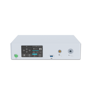
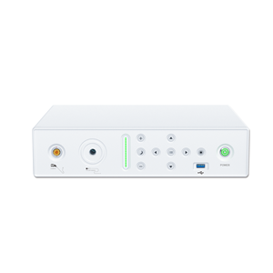
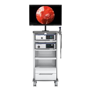
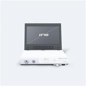
First, fully connect the laparoscope equipment, connect the laparoscope with the endoscope camera and the endoscopic camera system, turn on the pneumoperitoneum, high-frequency electrosurgical device, medical monitor, camera system, and cold light source in turn.
0.15mL/kg body weight dose of Dogmianbao was injected intramuscularly, and general anesthesia was given. After anesthesia, the test dog was placed on the operating table in a supine position, with a head high and a low tail, at an angle of about 20 degrees, and the tracheal intubation was connected to the monitor.
From the nipple to the front of the pubic bone, on both sides to the mid-axillary line, alcohol and iodine tincture are routinely disinfected. Put on a wound towel. The surgeon and assistant use two towel forceps to lift up the abdominal wall at the upper or lower edge of the umbilicus, and use wrist force to vertically penetrate the pneumoperitoneum into the abdominal cavity. Two breakthroughs in the fascia and peritoneum can be felt. If a 10mL syringe is connected to a negative suction or the saline in the syringe drops smoothly, it means that the puncture is correct and the abdominal organs are not injured, and carbon dioxide ventilation can be used. If the insufficiency is slow or fluctuating in breathing after the injection of normal saline, you can adjust the position of the pneumoperitoneum to move back and forth a few times. If you still see that the normal saline in the empty needle cannot drop smoothly, you should remove the needle and wear it again. In the process of inflating the abdomen, if the abdomen bulge is found to be asymmetrical, the needle should also be removed and re-pierced. When starting to inflate, the inflation speed should not be too fast, preferably 1L/min. When about 1L of carbon dioxide gas is injected, if the dog's blood pressure and heart rate are stable, it can be changed to high-flow inflation and the pneumoperitoneum pressure is maintained at 1.33kPa. Under the monitoring of the laparoscope, complete the entry of other cannula cones, and then insert the gallbladder grasping forceps and curved separating forceps from the puncture point. The serous membrane is stripped along the ampulla of the gallbladder to reveal the tapered part of the ampulla of the gallbladder, and then the cystic duct is separated along here, and the serosa is cut to free the cystic duct. When separating the cystic duct close to the common bile duct, especially when the cystic duct is too short, do not use electrocoagulation or resection to avoid accidental injury to the extrahepatic bile duct. Use an electrocoagulation hook to incise the posterior and lower serosal membrane of the cystic duct, insert a right-angle separation forceps from the top of the cystic duct and penetrate the fibrous connective tissue behind the cystic duct to completely free the cystic duct and confirm the relationship between the common bile duct and the cystic duct. Hold the titanium clamp to apply the clamp, place two titanium clamps at 0.5 cm from the common bile duct to close the cystic duct, and close the distal end of the cystic duct with one titanium clamp on the side of the neck of the gallbladder. Cut the cystic duct between the titanium clamps that clamp the cystic duct. Because the cystic artery is parallel to the cystic duct, it does not need to be separated separately and can be clamped together. The assistant uses the gallbladder grasping forceps to lift the distal end of the gallbladder and turn it upwards to fully expose the gap between the gallbladder and the gallbladder bed. Use an electrocoagulation hook to carefully separate the fibrous connective tissue between them, and properly grasp the depth of the stripping hook. Damage to liver tissue, causing hemorrhage on the liver surface, etc. The poking hole under the umbilicus or under the xiphoid process is the main outlet for removing the gallbladder and stones. Enter a specimen bag from the incision, open the specimen bag with grasping forceps, and then use grasping forceps to grasp the neck of the gallbladder or the titanium clip of the stump of the gallbladder duct, drag the gallbladder into the specimen bag, grab the mouth of the specimen bag, and put it in The inside of the cannula, along with the cannula and the gallbladder, are pulled out to the skin. Use a syringe to suck out the bile, gently rotate and pull to pull out the specimen bag.
After the gallbladder is taken out, the abdominal cavity is routinely closed with the incision, and the abdomen is inflated again. Carry out a careful examination of the abdominal cavity, especially the Calot triangle, duodenum and transverse colon, carefully observe the cystic duct and the stump of the cystic artery. If there is no bleeding in the gallbladder bed, the subhepatic space is clean, and the extrahepatic bile duct is not damaged, the operation can be ended. Although the gallbladder is accidentally punctured during the operation, the overflowing bile is fully flushed and sucked until the flushing fluid is clear and there is no active bleeding, and the drainage tube is not necessary to end the operation. If the surgical field is severely contaminated, the suction is not satisfactory even after washing, or there is suspected gallbladder bed oozing, biliary fistula, or effusion, a drainage tube should be placed in the subhepatic space.
After a comprehensive inspection of the abdominal cavity, slowly deflate the air. Be careful not to suddenly release the air. Pull out the other 3 cannulas except for the laparoscope under laparoscopic monitoring. When pulling out the laparoscopic cannulas, you should first enter the abdominal cavity slightly After pulling out the cannula, pull out the laparoscope. The 11mm puncture port is sutured with a conventional nodule, and the 5.5mm puncture port does not need to be sutured. The wound is disinfected with iodine tincture. Intravenous injection of Canine Xingbao resuscitated the dog.
When performing laparoscopic cholecystectomy, accurately identifying the anatomical characteristics of the cystic duct is a prerequisite to ensure the safe handling of the cystic duct during the operation. In the absence of a clear view of the anatomical relationship between the common hepatic duct, cystic duct and common bile duct, the cystic duct must not be rushed to avoid damage to the common hepatic duct or common bile duct.
Laparoscope is the most important surgical aid in general surgery. Laparoscopy technology has the characteristics of less trauma, accurate diagnosis and fast recovery. In the clinical diagnosis and treatment of small animals, veterinary laparoscopy is not only used as a diagnostic tool, but is now more used in minimally invasive surgery.


