Nasal Endoscope Is Used For Laryngo Examination And Treatment
The nasal endoscope is not only used for the inspection and surgery of the nasal cavity and sinuses, but also for the inspection and treatment of the nasopharynx, eustachian tube, etc. through the nasal cavity, sinuses and intracranial surgery. Let's learn about the use of nasal endoscope for laryngo examination and treatment.
Surgical methods:
For patients who have no contraindications for supporting laryngoscopy and general anesthesia, insert a supporting laryngoscope under general anesthesia to expose the glottis, insert a 0° or 30° nasal endoscope from the light source hole of the laryngoscope, with the lens bevel facing the affected side vocal cords After adjusting the lens until a clear and magnified image of the tissue is seen from the nasal endoscope, fix the body of the lens, and remove the diseased tissue under the microscope using conventional microlaryngosurgery methods. After the vocal cord lesions are removed, the nasal endoscope is sent into the subglottis from the light source hole of the self-supporting laryngoscope, and the subglottic area and the upper trachea are inspected (at this time, the 70° mirror can be replaced). If the lesion is found, it can be removed.
For those who are difficult to undergo support laryngoscopy and have contraindications to general anesthesia, cut the cricothyroid membrane under local anesthesia, and insert a 30° or 70° nasal endoscope through the cricothyroid membrane incision with the lens bevel facing the glottis. The lesion was completely resected under direct vision with a warped-head polyp forceps, and the neck incision was sutured routinely.
Nasal endoscopes have unique advantages for larynx examination and treatment:
1. Wide field of view, viewing angle up to 115°, bright light, and a certain magnification effect, clear imaging and high resolution;
2. The light can be refracted with the different oblique angles of the lens and can be rotated 360°. The larynx chamber, glottis, subglottic area and other parts can be inspected through the glottis or around the edge of the chamber. Treat lesions in areas that cannot be seen by the microscope;
3. For patients with contraindications to support laryngoscopy and general anesthesia, the combined application of cricothyrotomy and nasal endoscopy to remove benign tumors in the vocal cords and subglottic area can reduce incision and tissue damage, improve lighting conditions, and make The lesions are removed more thoroughly.
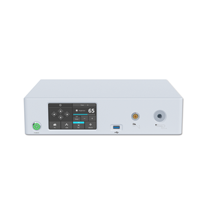
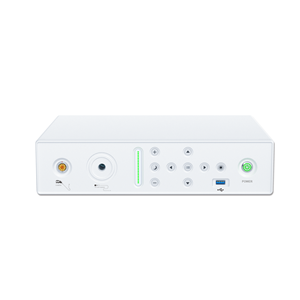
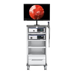
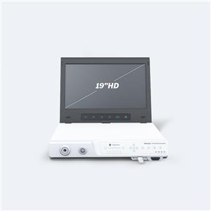
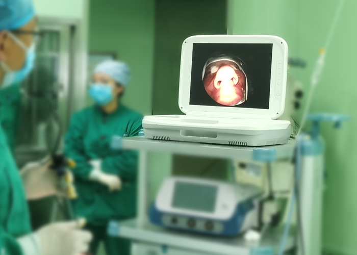
The nasal endoscope can be connected with the endoscope camera system, so that the operation method, operation cavity and other conditions can be completely displayed on the monitor, which is beneficial to the observation of the operation instructor, the surgeon, and the assistant.
- NEWS
- BLOG
- Industry News
- Company News

