【Gynecological endoscopy】Hysteroscopic endometrial resection
Endometrial resection is the use of hysteroscopy to remove or destroy the endometrium under the television to achieve the purpose of controlling bleeding. It is a safe and effective method to replace hysterectomy for the treatment of functional uterine bleeding.
Preoperative preparation
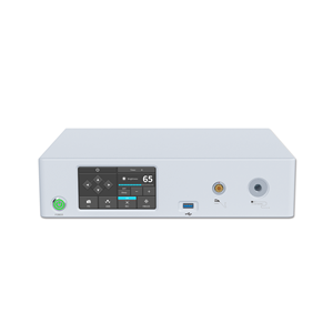
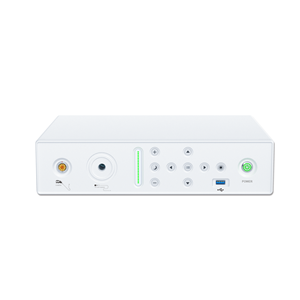
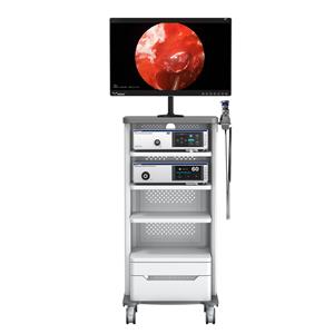
1. Prepare instruments and components: place the electrocautery device, endoscopic camera, monitor, cold light source, and multi-layer medical trolley in the operating room one day before the operation to ensure good performance. Electric cutting electrode, input water pipe, output water pipe, electric burning wire, cold light source wire, video conversion wire (except video conversion lens), put them in a formaldehyde fumigation box for 12 hours before use.
2. Prepare basic items: package and sterilize sterile forceps, uterine expansion rods, speculum, uterine probe, uterine curette, cervical forceps, large forceps and routine dressings for gynecological perineal surgery. Prepare 1500-3000ml of 5% glucose solution as uterine distention solution.
1. After the patient enters the room, an upper extremity venous access is established. After epidural block, the bladder lithotomy position was taken, and the height of the leg frame did not exceed 30cm. Place a cotton pad on the popliteal fossa and gently secure the knee to the leg brace with a bandage. The patient's legs were separated at an angle of 110-120°. Elderly patients are correspondingly smaller.
2. Routine perineal sterilization and laying sheets, correctly connect the wires and operating parts of each instrument, and turn on the power to keep it in working condition.
(1) Place the negative electrode of the electrocautery on the patient's muscle fullness and fully contact the skin to prevent burns. The general choice is the buttocks.
(2) Adjust the brightness of the cold light source to keep the brightness appropriate.
(3) Adjust the endoscope camera until the video screen image is clear, and wipe and disinfect the video conversion lens with 0.5% iodophor solution. First use sterile gauze dipped in iodophor solution to wipe the outer wall of the lens repeatedly for 3 minutes, and then wipe the iodophor solution with sterile dry gauze. It is confirmed by bacterial culture that the disinfection effect of this method is reliable. Iodophor solution wipe disinfection method also has the advantages of simplicity and speed.
4. Dilate the cervix and put it into the speculum. The nurse arranges the dilating rods in order from small to large. The donor gradually dilates the cervix until it can accommodate the outer sheath of the hysteroscope and puts it into the hysteroscope. During uterine expansion, closely observe the patient's consciousness, heart rate, blood pressure, respiration rate and blood oxygen saturation, and report any abnormality to the anesthesiologist and the surgeon in a timely manner.
5. Hang the hanging bottle-type disposable infusion set (referred to as hanging bottle) on the infusion stand, pour 500ml of 5% glucose solution as the dilatation fluid, connect the input water pipe and the output water pipe, and ensure the smooth perfusion and discharge of the dilatation fluid. Adjust the height of the infusion stand so that the liquid level in the hanging bottle is 1m higher than the operating table surface. Uterine distention pressure is maintained by using the liquid level difference of the distended fluid. The amount of perfusion is appropriate when the surgeon can clearly see the fundus and the opening of the fallopian tubes. 5% dextrose solution is a non-ionic solution and will not shock the patient during electrocution. The dilatation of the uterus can expand the uterine cavity, and the surgical field is clear and easy to operate. The amount of fluid in the uterine cavity maintains a dynamic balance. The uterine dilatation fluid flowing out of the efferent tube can not only take away the tissue removed by electric resection, but also lower the temperature of the uterine cavity, shrink local blood vessels, and reduce bleeding. The total amount of dilatation fluid is generally 1000-3000ml.
6. Under the monitoring of the video screen, the surgeon uses the electric cutting ring to cut the endometrium in turn, and then irons the cut surface with the spherical electrode. The circuit nurse adjusts the intensity of the electric burner according to the needs of the operator. The excised tissue fragments were sent for pathological examination.
7. After the electric resection is completed, connect the microwave therapeutic apparatus for adjuvant therapy.
After the operation, the hysteroscope and operating parts were rinsed with clean water, dried, and the metal joints were wiped with paraffin oil and stored for later use. The cold light source wire should not be folded to avoid damage to the light beam. The knobs of the electric burner, cold light source, endoscope camera and monitor are returned to the zero position and protected by a cover.


