How do animals undergo nasal endoscopy?
Animal endoscope examination and surgery can help relieve the pain of the animal and reduce the trauma of the animal owner’s mind. For example, a common nasal endoscope can diagnose various diseases of dogs and cats: such as runny nose, nosebleeds, chronic sneezing, suspected foreign bodies, and obvious pathological changes visible by X-rays. So how do animals undergo nasal endoscopy?
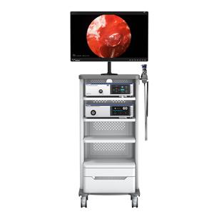
Common animal nasal endoscopes are rigid or fiber optic. The rigid lens can provide the best optical image, while the soft lens can be used for posterior nasal examination (the soft lens is inserted into the nasopharynx through the oral cavity to see the posterior nasal cavity). The rigid lens can be used for biopsy and foreign body removal. The rigid lens also has Working channel and has flushing or suction function.
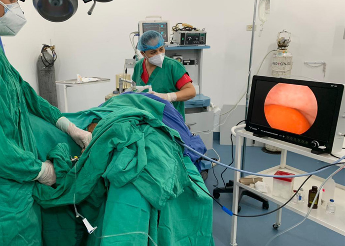
Animal nasal endoscopy:
Nasal endoscopy needs to be performed under general anesthesia of the animal, but due to the dense nerves in the nasal cavity, it is extremely sensitive, even if the animal has reached the depth of anesthesia to the degree of surgery, touching the mucosa in the nasal cavity may cause the animal to sneeze and sudden heart rate Accelerate pain reactions such as increased blood pressure, so it is necessary to apply systemic pain relief and local anesthesia to all animals under nasal endoscopy. In addition, massive bleeding in the nasal cavity is often seen during the operation of nasal endoscopy, so coagulation-related inspections must be performed before nasal endoscopy. Normal temperature normal saline can be used to fully rinse during the operation to ensure a clear inspection field. It can also avoid the introduction of a large number of bacteria to prevent the growth of bacteria, and it can also wash away the blood clots and cell debris on the surface of the tumor, which may affect the inspection, and expose the diseased area.
Frontal inspection of nasal cavity:
For inspection of the anterior part of the nasal cavity with a rigid lens, first perform the normal side exploration, and then explore the side nasal cavity;
Nasopharyngeal examination
Use a soft lens to explore the nasopharynx with a "J"-shaped retrograde method;
Regardless of whether you use a rigid or soft lens, you must insert it gently and slowly to ease the pain.
- NEWS
- BLOG
- Industry News
- Company News

