【ENT Endoscopy】Nasal Septum Correction under Nasal Endoscope
[ENT Endoscopy] Nasal Septum Correction under Nasal Endoscope
Surgical equipment and instruments:
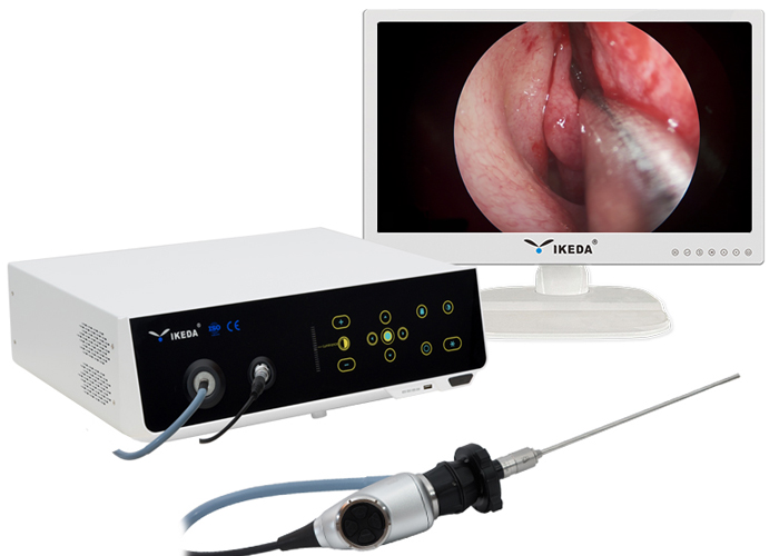
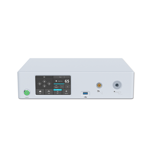
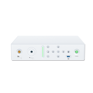
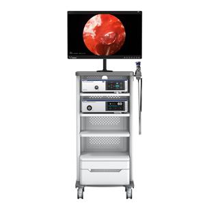
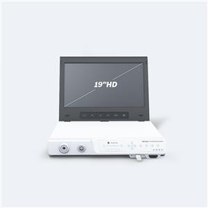
Endoscope camera, cold light source, medical monitor, 0° nasal endoscope and straight ethmoid rongeur, nasal septum spreader, suction device, all strippers and rongeurs for traditional nasal septum correction.
Surgical methods:
First, connect the endoscope camera, cold light source, medical monitor, and 0° nasal endoscope, do white balance, and check whether the machine is operating normally. Then the patient lies on his back, with shoulder pads, and his head slightly tilted towards the surgeon. Routinely sterilized sterile towels. Local anesthesia was used. The nasal mucosa was surface anesthetized with 1% dicaine cotton pad, and the incision was anesthetized with 2% lidocaine infiltration. The entire operation was completed under a 0°endoscope. After local anesthesia, carefully check under a 0°endoscope to see the deviated part, shape and scope. Conventional incisions are sufficient to ensure the separation of the mucoperiosteal flap underneath the perichondrium and periosteum. Separate upward to the upper edge of the nasal septum, rearward to the back of the deflection, and downward to the bottom of the nose. During the separation, the deflection bone can be removed with cartilage forceps and bone forceps in stages until the deflection is completely corrected. After the operation, the nasal cavity was filled with petroleum jelly gauze and removed after 2 days. The mucosal incision is covered with a small piece of soluble hemostatic adhesive to reduce the bleeding of the mucosal incision after the operation and promote the healing of the wound. For patients with sinusitis or nasal polyps, endoscopic sinus surgery or nasal polyp removal can be performed at the same time. For patients with nasal adhesions, the adhesive tapes were removed at the same time.
The nasal septum is deflected to one or both sides or locally formed protrusions, causing nasal cavity and sinus dysfunction and producing symptoms, which can be diagnosed as nasal septum deviation. The high deviation of the nasal septum is one of the important reasons for the occurrence and recurrence of sinusitis. The traditional submucosal resection of the nasal septum is affected by the patient's position and frontal mirror illumination. The deviation of the posterior high position and the posterior segment is not corrected, resulting in recurrent sinusitis and repeated bleeding. Under the nasal endoscope, the whole picture of the high and posterior segment of the nasal septum can be seen very clearly, thereby providing the possibility to completely correct the deflection; under the nasal endoscope, the anterior posterior ethmoidal nerve and the medial innervation of the posterior superior nasal nerve The mucosal anesthesia site in the area is more accurate and the effect is more significant; the nasal endoscopy has sufficient lighting, and can be deepened at any time with the continuous separation of the periosteum and periosteum, so as to fully expose the high and posterior deflection cartilage and Bone, which can safely and completely correct the deviation, reduce the recurrence of sinusitis and nasal polyps, and also prevent the collapse of the nasal bridge and the perforation of the posterior segment of the nasal septum.
The correction of nasal septum under nasal endoscope is operated under direct vision, with a clear vision and able to see all parts of the nasal cavity. It overcomes the shortcomings of insufficient illumination of the traditional operation field and the need to explore the deep parts. In addition, the endoscope can directly reach the deep part, so that the deviation of the high position and the posterior segment can be easily corrected.Therefore, the endoscope is better suitable for various types of nasal septal deflection surgery, especially the high and posterior nasal septum. Deviation.
Under the clear field of vision of the nasal endoscope, it can accurately separate the periosteum and periosteum, especially the separation of the mucoperiosteum at the nasal septal spine and ridge, which can prevent the tear of the nasal mucosa and avoid the perforation of the nasal septum.
For cases of spines and ridges that are not accompanied by deflection, they can be simply removed to reduce trauma and facilitate postoperative recovery. The operation is delicate and the mucosal damage of the lateral wall of the nose is small, so as to avoid the formation of postoperative nasal cavity adhesion.
For patients with deviated nasal septum combined with nasal polyps and/or sinusitis, this operation can fully improve the visual field of endoscopic sinus surgery, and these two operations can be performed at the same time, which not only shortens the hospital stay, but also saves patients The cost, avoiding the pain of multiple operations, is very popular with patients.
