Removal Of Cholesteatoma In External Auditory Canal Under Ear Endoscope
External auditory canal cholesteatoma is a common clinical disease, which can cause compressive bone destruction. It also produces a proteolytic enzyme that promotes osteolysis and causes the bony external auditory canal to corrode and expand, which is sac-shaped to the external auditory canal. Extension of the walls and upper tympanum, or destruction of the posterior wall of the external auditory canal to invade the mastoid, in addition to directly affecting the patient’s hearing, severe cases can also cause intracranial and extracranial complications.
Clinical manifestations of cholesteatoma of the external auditory canal: ear fullness, earache, otorrhea, tinnitus and hearing loss.
Removal of cholesteatoma in external auditory canal under ear endoscope
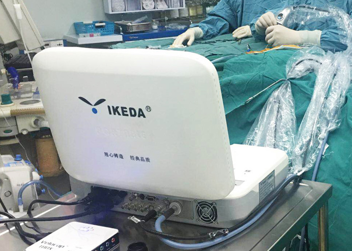
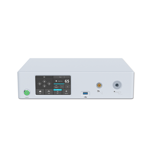
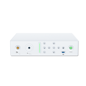
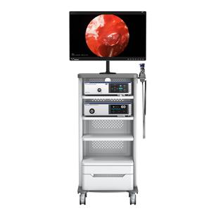
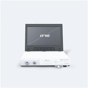
Surgical equipment: hard tube Ear Endoscope, Endoscope Camera System, Cold Light Source, Medical monitor, surgical instruments, etc.;
Surgical methods:
The patient was placed in a supine position, the affected ear was deflected to the opposite side by 45°, and routine disinfection and draping were performed. Under the ear endoscope, the cholesteatoma-like material was gradually separated and removed along the external auditory canal with a stripper. Those with granulation tissue hyperplasia were removed with cup-shaped polyp forceps. Probe the destruction of the bone wall of the external auditory canal under a 30° ear endoscope, clean up necrotic bone fragments, cholesteatoma-like materials, granulation tissue and pus, and try not to damage the skin of the external auditory canal. After thoroughly removing the lesion, apply a layer of erythromycin ointment to the external auditory canal cavity. Oral antibiotics were taken for one week after the operation, and the operation cavity was checked under an ear endoscopy on 1, 2, 3, 7, 14, and 28 days after the operation, and regular rechecks for more than 1 year thereafter.
Comparison of treatment methods:
Traditional external auditory canal cholesteatoma surgery is generally performed under frontal mirror or headlamp illumination, especially for children. Under such illumination, the surgical field is narrow, the light is weak, and the ear canal is poorly exposed, so the operation is relatively difficult; under the microscope Surgery for cholesteatoma of the external auditory canal is complicated and expensive, and it is difficult to purchase it in primary hospitals. Ear endoscopy under direct vision for external auditory canal cholesteatoma surgery can be directly inserted into the external auditory canal and close to the tympanic membrane, so that the surgical field can be bright, and the relationship between the lesion and the structure of the external auditory canal and middle ear can be observed, and complications can be avoided to the greatest extent. The operation is simple and convenient, and the medical monitor is connected, and the operation is explained at the same time, which is extremely beneficial to clinical teaching. Ear endoscopy for external auditory canal cholesteatoma surgery, with good illumination view and clear magnified imaging, the relationship between the disease and the middle ear and other important structures can be observed from different angles. In the case of the lack of funds in the basic hospital to purchase an operating microscope, the ear Speculum is the preferred alternative device. However, cholesteatoma surgery of the external auditory canal under the ear endoscope also has its own shortcomings: (1) Because the displayed image is a two-dimensional image, it has no stereoscopic effect, and it is difficult for beginners to adapt; (2) The operation is one-handed operation. Further increase the difficulty of the operation.
The ear endoscope can provide good illumination, magnification and high-resolution images. At the same time, the endoscope with multiple viewing angles can bypass physiologically curved anatomical structures or blocking structures to detect blind areas that cannot be seen directly, and can be very close to the disease. Make careful observations.
