Application Of Veterinary Laparoscope In Animal Reproduction
With the continuous improvement of veterinary endoscope technology, veterinary laparoscopy has been widely used in the diagnosis and surgical treatment of veterinary clinical animal diseases, as well as animal husbandry and embryo transfer, especially in animal reproduction.
Application of veterinary laparoscope in animal reproduction
1. Application in artificial insemination
(1) Identification of estrus
After the doctor puts the laparoscope into the abdominal cavity of the animal through the cannula, install the air delivery tube on the air delivery valve of the cannula, and first send in a small amount of gas, connect the optical cable to the optical cable interface of the lens, turn on the power switch, and observe whether the objective lens is through the eyepiece In the abdominal cavity, after confirming that it is correct, continue to ventilate into the abdominal cavity until the stomach and intestines move forward, and space appears in the back of the abdominal cavity and pelvic cavity. After the reproductive organs are clearly seen, stop the air supply and observe. During observation, the distance between the observed organ and the objective lens and the direction of the lens should be changed at any time to obtain a good observation angle and line of sight. The size and shape of the ovary, the size of the follicle, and the presence or absence of the corpus luteum should be observed from the right side. After observing the right side, observe the left side again. When observing the ovary, it is observed that the volume of the follicle reaches the maximum, the blood vessels in the mesangium are very thick, the diameter of the follicle can reach 0.5~1.0cm, the follicle is spherical protruding from the surface of the ovary, the wall of the follicle is very thin, and the follicle is filled with translucent dark red follicles Liquid, it can be determined that the animal is in estrus.
(2) Artificial insemination
①Punch and inflate: First, punch a hole on the left side of the midline of the lower abdomen with a 7mm cannula and probe, put it in the inflation tube, open the valve regulator and inflate it, close the valve regulator after the abdomen is inflated, and take it out The inflatable tube is put into the laparoscopic tube to supply the intra-abdominal light source. Then insert a 5mm cannula and probe on the right side of the midline of the lower abdomen. This tube is used to find the uterine horn with the help of a light source.
②Look for the uterine horn: The uterine horn is just in front of the bladder. If the bladder is enlarged, a laparoscopic tube or a 5mm cannula can be used to pull it aside. A surgical cannula and forceps can be inserted into the 5mm cannula to help determine the position of the uterus. Take care not to damage the internal organs. The visceral fat sometimes covers the uterus. You can use a laparoscopic tube to pull it apart until you find the uterine horn.
③Insemination: prepare a straw, first draw 0.3ML of gas, and then draw the required semen. After the doctor finds the uterine horn, draw out the probe from the 5mm cannula and insert the straw, taking special care not to bring contaminants into the abdominal cavity. The tip of the straw can be inserted into one side of the uterine horn through a laparoscope, and the exact position should be between the bifurcation of the uterine horn and the uterine horn. Use a syringe to inject semen, with the help of a light source, you can clearly see the semen enter the uterine horn through the straw. Withdraw the straw, then repeat the above process on the other side of the uterine horn, and infuse again.
The use of laparoscopy for insemination can greatly increase the conception rate during estrus, which has great breeding value and production significance for the breeding and expansion of sheep.
2. Application in pregnancy diagnosis
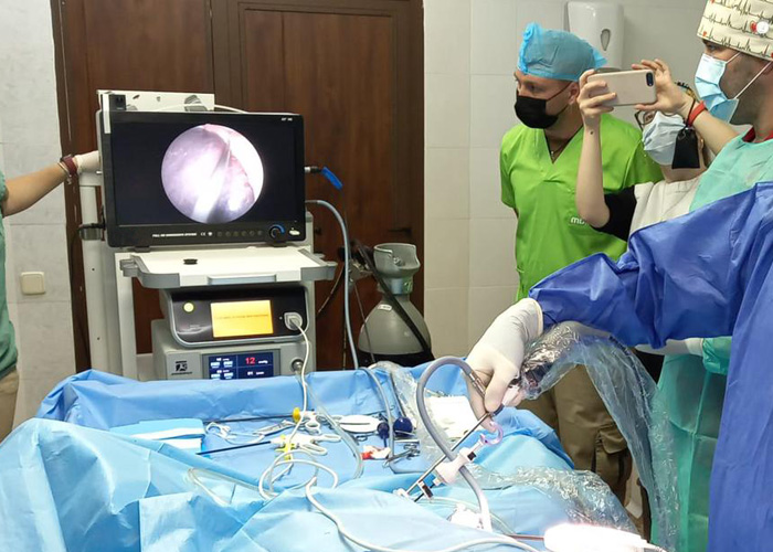
During operation, fix the ewe on a mobile operating rack, the surgical site is 8~10cm in front of the breast, punch the punch into the abdominal cavity, and infuse an appropriate amount of carbon dioxide gas through the punch to separate the abdominal wall from the internal organs. Then observe the development of the ewe's uterus with the help of the light source and lens of the laparoscope. If a significant difference in the size of the uterus on both sides is observed, and the larger uterus or both uteruses (twin or multiple pregnancies) have slight peristalsis, and the uterine surface is rich in blood vessels, it can be determined as pregnancy.
3. Application in embryo transfer
① Observation of the effect of superovulation: Use laparoscopy to observe the uterine state and ovarian response of the embryo transfer donor and recipient sheep, thereby increasing the success rate of embryo recovery, reducing ineffective operations and surgical damage to the donor and recipient, and shortening the embryo Recycle to the time of transplantation.
② Observation of estrus at the same time: After using reproductive hormones on animals, estrus at the same time occurs. At this time, laparoscopy can be used to examine the donor and recipient sheep to determine the development of follicles and the presence or absence of corpus luteum. When the recipient sheep has a functional corpus luteum on its ovary, it can be used as a transplant recipient.
③ Embryo collection: After superovulation, observe the state of the uterus and ovarian response with a laparoscope, and use the guidance of the laparoscope to collect embryos. After flushing one side, flush the other side. This method is simple, fast, and convenient. High efficiency and little damage to animals.
④ Embryo transfer: Using a laparoscope and supporting operating equipment, the recipient sheep is lying on its back on a restraining frame, in a front-low-back posture, and it is sheared and disinfected according to the routine operation. Use a punch with a trocar about 5cm in front of the breast and 4~6cm on both sides of the midline of the abdomen to make a hole. Insert the laparoscopic tube and probe on the left and right sides (the front end is blunt). Observe the number of corpus luteum and determine the grade under the monitoring of laparoscope. Use oval forceps to clamp the junction of the uterine tube and pull it out of the abdominal wall. Push the embryo in with a transfer needle with a 1ML syringe, and then return it to the abdominal cavity for repositioning. The biggest advantage of the laparoscopy method compared with the surgical method and the small picking method is that the ovaries and corpus luteum of the recipient sheep are very little touched, the fixation and traction of the uterine tube junction are slight, and the embryo development and implantation are generally not adversely affected. Affected, adhesion will not occur.
