Application of rigid tube nasal endoscope in throat examination and operation
Rigid tube nasal endoscope is an otolaryngology device that is an optical device that can perform detailed inspection of the nasal cavity. Connected with cold light source, endoscopic camera and monitor, it can go deep into the nasal cavity, so that all pathological changes hidden in the nasal cavity can be clearly displayed, not only can effectively treat rhinitis, sinusitis, nasal polyps, nasal septum deviation and other nasal diseases , It can also be used to check and operate the throat under the supporting laryngoscope. Get to know it together.
Application of rigid tube nasal endoscope in throat examination and operation
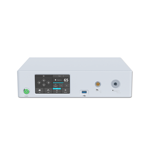
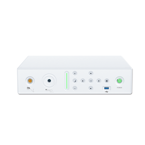
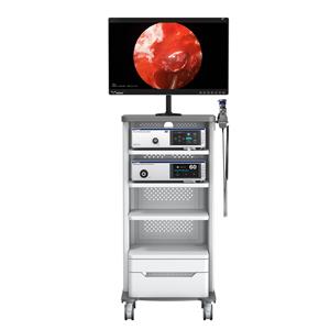
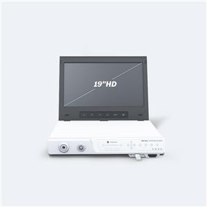
Equipment: nasal endoscopes, supporting laryngoscopes, endoscopic cameras, medical monitors, medical trolleys, etc. with viewing angles of 0°, 30°, 70°;
Surgical methods:
Under general anesthesia, insert the supporting laryngoscope to expose the glottis, insert the 0° or 30° nasal endoscope from the light source hole of the laryngoscope, with the lens bevel facing the affected side vocal cords, adjust the lens until it is seen from the nasal endoscope or monitor After a clear and magnified image of the tissue, the scope is fixed, and the diseased tissue is excised or cut off from the junction of the diseased tissue and the normal tissue with a laryngoscope or laryngeal scissors. After the vocal cord lesions are removed, the nasal endoscope is sent into the subglottis from the self-supporting laryngoscope tube. If it is normal, the nasal endoscope and the supporting laryngoscope are withdrawn. For those with difficulty in intubating the supporting laryngoscope and contraindications to general anesthesia, after the cricothyroidectomy under local anesthesia, a 30° or 70° nasal endoscope is inserted through the incision, with the lens bevel facing the glottis, and then tilted The head polyp forceps are inserted into the subglottic area from the incision, and the lesions are removed under direct vision of the endoscope.
In traditional laryngeal microsurgery, the microscope can only provide light along the vertical axis. Therefore, the lesions in the anterior vocal cord, laryngeal chamber, and subglottic area are easily overlooked, missed, or difficult to operate, and it is not easy to achieve satisfactory results. In laryngeal examination and surgery, the nasal endoscope can not only replace the microscope, but also has its unique advantages: a wide field of view, a viewing angle of up to 115°, bright light, and can meet the lighting needs of various depths of the larynx . And it has non-focus performance, the imaging is very clear from one millimeter to several hundred millimeters, and there is a certain degree of magnification. With high resolution, it is easy to distinguish the boundary between diseased tissue and normal tissue, making the operation more delicate. Secondly, the light can be refracted with the different oblique angles of the lens. The lens body can rotate 360°. It can be used to inspect the "blind areas" such as the larynx chamber and subglottic area that cannot be seen by the microscope through the glottis or bypass the edge of the chamber. And treatment. On the other hand, for patients who are contraindicated to use support laryngoscope and general anesthesia, when using transcricothyrotomy to remove benign tumors in the vocal cords and subglottic area, the combined application of nasal endoscope can reduce the incision and tissue damage. Improve the lighting conditions, so that the lesions can be removed more thoroughly.


